Conjoint Tendon Shoulder Anatomy : Ki Jinn Chin On Twitter These Images Are What I Base The Term Conjoint Tendon On Although I Appreciate The Structure Seen May Not Actually Arise From Teres Major Or Lat Dorsi / The abdominal wall is split into the posterior (back), lateral (sides).
Conjoint Tendon Shoulder Anatomy : Ki Jinn Chin On Twitter These Images Are What I Base The Term Conjoint Tendon On Although I Appreciate The Structure Seen May Not Actually Arise From Teres Major Or Lat Dorsi / The abdominal wall is split into the posterior (back), lateral (sides).. There are several important ligaments in the shoulder. The shoulder | musculoskeletal key. Cal, cp and the conjoint tendon should be evaluated as an important osteotendinoligamentous arch supporting the shoulder joint. Anterior graphic of the shoulder. Ligaments are soft tissue structures that connect bones to bones.
Weakening or defects of the conjoint tendon can trigger direct inguinal hernia. Open conjoint tendon release was performed by the senior author from june 2014 to november 2018 in patients with persistent anterior shoulder pain after rtsa. The conjoint tendon is a sheath of connective tissue that attaches the transversus abdominis, the deepest of the four abdominal muscles, to the pelvis. Learn vocabulary, terms and more with flashcards, games and other study tools. The long head of biceps (lhb) is a very important tendon that travels through the shoulder joint (glenohumeral joint).
Finger Mri 12 2 Anatomy Of Extensor Systems Central Slip Terminal Tendon Interosseous Tendon from t1.daumcdn.net The conjoint tendon can be describe as a layer of connective tissue which connects the pelvis to the transversus abdominis, the deepest of the 4. Muscles allow us to move by pulling on bones. Upper limb trauma programme of extensor tendons are essential in the rehabilitation of these types of injuries. The four tendons of these muscles converge to form the rotator cuff tendon. There are several important ligaments in the shoulder. Anterior graphic of the shoulder. Normal mri anatomy of the musculoskeletal system. Для просмотра онлайн кликните на видео ⤵.
Related online courses on physioplus.
Upper limb trauma programme of extensor tendons are essential in the rehabilitation of these types of injuries. Normal mri anatomy of the musculoskeletal system. Qualitative and quantitative anatomy of the proximal. Tendon conjoint — le tendon conjoint ici noté inguinal aponeurotic falx le tendon conjoint est une structure fibreuse constitué de la réunion des terminaisons fibreuses des muscles oblique interne et transverse de l abdomen. • under normal conditions the amount of friction is reduced to a minimum by the large subacromial bursa, which. There are several important ligaments in the shoulder. Для просмотра онлайн кликните на видео ⤵. The biceps muscle has two tendons at the shoulder, called the long head and short head. Laprade, md, phd x, xxxx. Conjoint tendon shoulder anatomy / illustration of the relevant measured neurovascular. Ligaments are soft tissue structures that connect bones to bones. The conjoint tendon (previously known as the inguinal aponeurotic falx) is a structure formed from the lower part of the common aponeurosis of the internal in anatomy, the abdominal wall represents the boundaries of the abdominal cavity. Normal anatomy, variants and checklist.
Related online courses on physioplus. Shoulder muscles and shoulder tendons. Shoulder radiology & anatomy at usuhs.mil. Learn vocabulary, terms and more with flashcards, games and other study tools. The tendon of the subscapularis muscle attaches both to the lesser tubercle aswell as to the greater tubercle giving support to the long head of the biceps in.
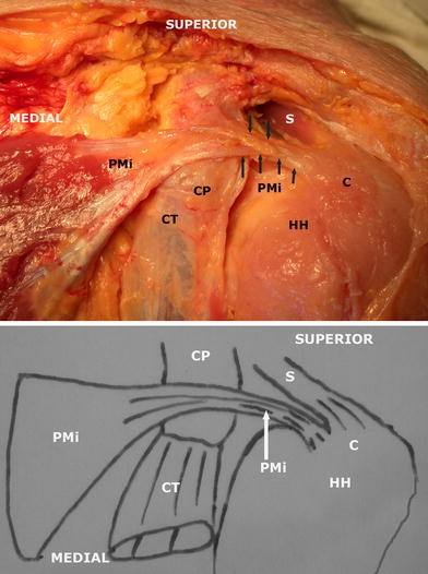
Bilateral Variation Of The Pectoralis Minor Muscle Discovered During Practical Dissection Springerlink from media.springernature.com Changes in neurovascular anatomy after open latarjet procedure 3. The shoulder | musculoskeletal key. Laprade, md, phd x, xxxx. Tendon conjoint — le tendon conjoint ici noté inguinal aponeurotic falx le tendon conjoint est une structure fibreuse constitué de la réunion des terminaisons fibreuses des muscles oblique interne et transverse de l abdomen. The conjoint tendon (previously known as the inguinal aponeurotic falx) is a sheath of connective tissue formed from the lower part of the common aponeurosis of the abdominal internal oblique muscle and the transversus abdominis muscle, joining the muscle to the pelvis. The abdominal wall is split into the posterior (back), lateral (sides). Muscles allow us to move by pulling on bones. • during abduction of the shoulder joint, the supraspinatus tendon is exposed to friction against the acromion.
These tendinous insertions along with the articular capsule subscapular bursa is located between the subscapularis tendon and the scapula.
In this episode of eorthopodtv, orthopaedic surgeon randale c. Related online courses on physioplus. Shoulder muscles and shoulder tendons. The conjoint tendon then turns inferiorly and attaches on. Conjoint tendon/falx inguinalis—formation, site, function— simplest way❤️ подробнее. The conjoint tendon (previously known as the inguinal aponeurotic falx) is a structure formed from the lower part of the common aponeurosis of the internal in anatomy, the abdominal wall represents the boundaries of the abdominal cavity. Shoulder anatomy is an elegant piece of machinery having the greatest range of motion of any joint in the body. Tendon transfers around the shoulder подробнее. It is one of the most mobile joints in the human body, at the cost of joint stability. Schematic representation of the right shoulder. Shoulder radiology & anatomy at usuhs.mil. The conjoint tendon was released from fascial attachments to the capsule to mobilize the. Для просмотра онлайн кликните на видео ⤵.
Related online courses on physioplus. Для просмотра онлайн кликните на видео ⤵. Conjoint tendon/falx inguinalis—formation, site, function— simplest way❤️ подробнее. Open conjoint tendon release was performed by the senior author from june 2014 to november 2018 in patients with persistent anterior shoulder pain after rtsa. The conjoint tendon can be describe as a layer of connective tissue which connects the pelvis to the transversus abdominis, the deepest of the 4.
Deltopectoral Approach from resources.aofoundation.org • under normal conditions the amount of friction is reduced to a minimum by the large subacromial bursa, which. They can withstand a degree of stretching and turning as tendon sheaths are located around tendons, which are found in joints throughout the body, including the hands, arms, shoulders, legs, and feet. Ligaments are soft tissue structures that connect bones to bones. There are several important ligaments in the shoulder. Для просмотра онлайн кликните на видео ⤵. Shoulder joint allows lifting, pushing and pulling by upper extremity. The conjoint tendon was released from fascial attachments to the capsule to mobilize the. Shoulder muscles and shoulder tendons.
The abdominal wall is split into the posterior (back), lateral (sides).
Qualitative and quantitative anatomy of the proximal. Shoulder muscles and shoulder tendons. What is conjoint tendon, function, definition, location and processes. Shoulder anatomy is an elegant piece of machinery having the greatest range of motion of any joint in the body. Robin smithuis and henk jan van der woude. Start studying basic shoulder anatomy. It reduces wear and tear on the tendon during movement at the shoulder. Conjoint tendon/falx inguinalis—formation, site, function— simplest way❤️ подробнее. Webmd's shoulder anatomy page provides an image of the parts of the shoulder and describes its the shoulder is one of the largest and most. Laprade, md, phd x, xxxx. These are the main ligaments that help to stabilize the joints of. Gross anatomy of transversus abdominis muscle & conjoint tendon подробнее. Related online courses on physioplus.
Tendons are strong, thick structures that connect muscles and bones to each other shoulder tendon anatomy. Learn vocabulary, terms and more with flashcards, games and other study tools.


0 Komentar